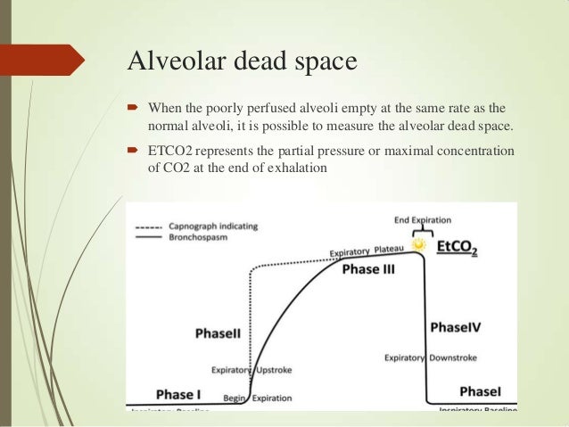
Decreased cardiac output or lung perfusion increases dead space and makes ventilation less efficient. Unlike anatomic dead space, which is fixed, physiologic dead space can change from minute to minute with changes in cardiac output and pulmonary blood flow. Physiologic dead space, consists of alveoli that are ventilated but lack capillary blood flow to pick up oxygen and drop off carbon dioxide. It’s called anatomic because it’s fixed by anatomy and doesn’t change. Since it is not perfused, oxygen can’t be absorbed and carbon dioxide cannot be eliminated- that air here is wasted breath. It consists of conducting airways such as the trachea, bronchi, and bronchioles -structures that don’t have alveoli. Types of Dead Space Anatomic Dead SpaceĪnatomic dead space consists of the fixed parts of the respiratory tract that are ventilated but not perfused. There are three types of dead space: anatomic, physiologic, and the dead space belonging to any airway equipment being used to assist ventilation. What Is Dead Space?ĭead space is the portion of the respiratory system where tidal volume doesn’t participate in gas exchange. This article discusses the concept of dead space and it’s clinical use in recognizing hypoventilation and preventing hypoxia and hypercarbia. Having a tidal volume close to, or smaller than the patient’s dead space can lead to significant hypercarbia, hypoxia, and respiratory failure. A more subtle sign is that tidal volume becomes shallower. The most obvious sign is slowing of the rate of breathing. Hypoventilation from sedation, pain medications, anesthesia in the immediate postoperative period is common. Ventilation may exceed perfusion in parts of the lung resulting in increased physiological dead air space.Understanding anatomic dead space is important to recognizing subtle hypoventilation.

Air may reach the periphery of the lungs but fail to make contact with the capillary blood. The alveoli become permanently damaged (see video above).This is why breathlessness and fatigue are common symptoms of COPD. This extra effort can make the patient feel very tired. However, this does not mean that your oxygen levels are low because the breathing muscles around the chest are working harder to compensate.


As the lungs become hyper-inflated they elongate and flatten, which means the diaphragm does not work as well as it should. As a result, air gets trapped in the lungs and the lungs get bigger (hyper-inflated). These changes cause the air sacs (alveoli) to close before you have fully exhaled.

In emphysema, exposure to an irritant over many years causes an inflammation in the lungs which causes the following changes: Please note there is no audio for this animation


 0 kommentar(er)
